-
熔体包裹体,又称熔融包裹体,为岩浆体系中矿物结晶生长时所捕获的微量硅酸盐熔体(Sorby, 1858)。熔体包裹体保存着原始岩浆和初始热液的物质组成及物化条件信息,成为反映岩浆-热液演化和成岩-成矿环境的直接证据(Davidson et al., 2006; Zajacz et al., 2007; Esposito et al., 2014; Hamada et al., 2020)。自20世纪以来,对矿物内部熔体包裹体进行测试和分析的技术手段迅速发展,吸引着地质学、地球化学、分析化学等专业学者的关注,并结合相关科学问题进行了大量的应用研究(Audétat et al., 2014; Cannatelli et al., 2016;张道涵等, 2017;王国光等, 2020)。
早期学者对熔体包裹体的研究侧重于对其特征的观察和描述,并初步提出了熔体包裹体可以作为探讨火成岩起源的潜在工具的设想(Zirkel, 1873)。然而,受制于当时分析技术的落后,多相熔体包裹体难以准确识别,导致其在地质学研究受限,进而落后于“同族”流体包裹体的应用。百年之后,Barrabé和Deicha首次尝试通过高温加热对石英中的熔体包裹体进行了均一化实验,获取了熔体包裹体的物化信息,使得该方法发展为获取包裹体内部信息的关键技术(Cannatelli et al., 2016)。至1970年前后,地球化学学家开始利用电子探针对熔体包裹体展开成分测试,进而推动了熔体包裹体在火山岩成因方面的研究(Sobolev et al., 1975)和月球样品的分析(Roedder et al., 1970)。同期,部分学者专注于探索熔体包裹体的起源和研究方法(Clocchiatti, 1975; Roedder, 1979),极大地推动了熔体包裹体研究的发展。到20世纪90年代初期,地质学家将熔体包裹体研究重点放在了火山系统的应用,讨论了它们在确定岩浆喷发前挥发性成分中的作用(Roedder, 1992; Johnson et al., 1994; Lowenstern, 1994; 1995; Sobolev, 1996)。随着微区原位分析技术的发展,熔体包裹体研究成果呈指数增长,尤其是单个熔体包裹体显微测温和成分分析技术的发展,加速了成矿岩体的熔体包裹体研究进程,对岩浆热液矿床的成因提出了新认识(王蝶等, 2017)。虽然,针对熔体包裹体的探索已经历了近百年的历程,但仍然存在一系列问题,成为新的研究难点和热点,例如熔体包裹体成分的变化、边界层效应及捕获后的改造等(Wallace et al., 2021)。
随着宜居地球这一重大科学问题的提出,以地球系统科学思维探讨深部过程与浅部效应耦合的研究方兴未艾(朱日祥等, 2021)。而作为宜居地球深部过程的重要见证者和记录者——熔体包裹体,具备了新的使命。因此,深入探讨熔体包裹体分析方法的研究进展和存在问题成为重中之重。本文针对熔体包裹体分析的相关测试流程、技术手段和研究进展进行了系统总结,希望对相关学科的科研工作者了解并应用熔体包裹体研究提供帮助。
2熔体包裹体岩相学观察2.1熔体包裹体的厘定开启熔体包裹体研究的第一步也是最重要的一步就是有效识别熔体包裹体及其组合(Cannatelli et al., 2016)。然而,由于在成岩-成矿作用早期形成的熔体包裹体数量较少,并且大都在后期发生一定程度的改造,形成较为复杂的内部结构,使之和流体包裹体相比,观察起来更为困难。因此,精细厘定熔体包裹体的类型是首要任务。
目前对于熔体包裹体的分类主要依据2个原则。其一是按照熔体包裹体的捕获时间可以分成原生、次生和假次生包裹体3种类型(Roedder, 1979)。原生包裹体就是在矿物生长的同时被捕获于主矿物生长缺陷中的高温岩浆熔体。它们的直径较大,通常随机独立分布在主矿物中,是准确获取岩浆演化信息的有效载体。次生包裹体是指在主矿物晶体形成之后被捕获于裂隙等晶体缺陷中的包裹体。由于其无法代表成岩-成矿作用早期或同期的熔/流体成分,因此在进行镜下观察时要对这些包裹体进行筛选。这类熔体包裹体往往具有体积小、成群成带分布的特点。假次生包裹体和次生包裹体的捕获机制类似,但捕获后主矿物会沿其上部晶面继续生长,成为硅酸盐熔体演化过程的重要记录者,因此假次生熔体包裹体也记录了部分“原生”岩浆信息。
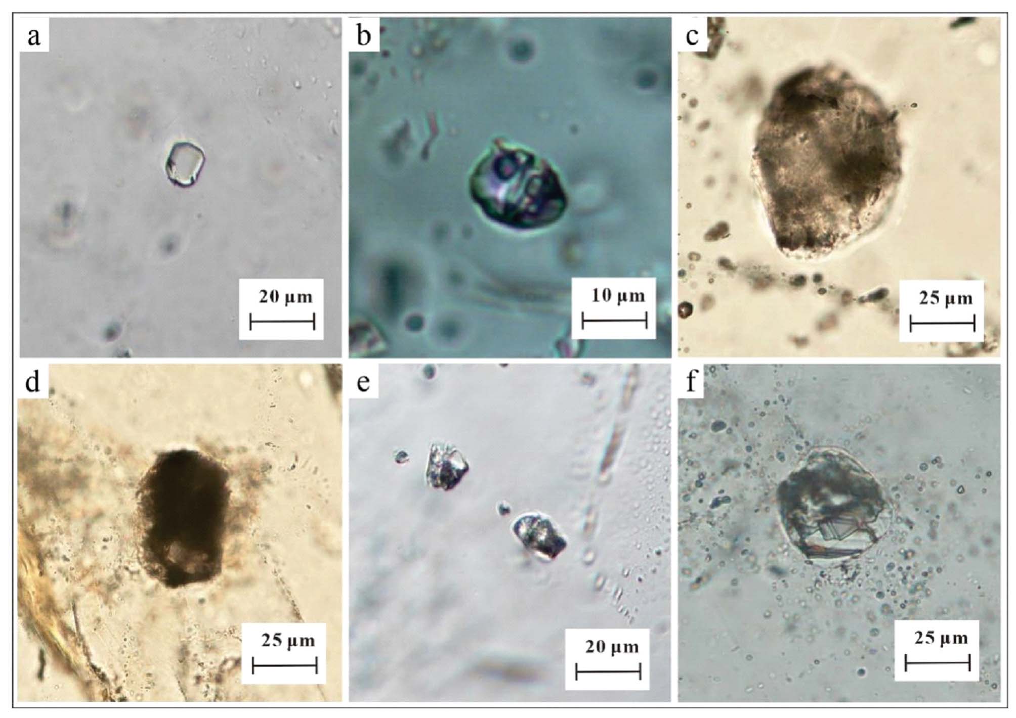
另一种分类方法是根据包裹体内部结构可以分为玻璃质、结晶质和熔-流体包裹体(图1a~f)。最初捕获的原始岩浆的熔体包裹体显示完全玻璃质的组成,但由于主矿物在岩浆通道中快速上升会经历一个淬火过程,诱发玻璃质熔体包裹体内部出现一个或多个气泡;而随着冷却速率减慢,包裹体内部还会逐渐发生细小结晶;当冷却速度到达一定程度时,包裹体完全结晶,内部呈现网状结晶相(王蝶等, 2017)。结晶质包裹体因其内部成分不均匀,所以对其做成分分析前通常要进行均一化实验。熔-流体包裹体是指硅酸盐熔体与流体(水或CO2)相同时被圈闭的一类包裹体(常海亮等, 1998),因此该类包裹体是研究岩浆-热液过渡阶段流体演化的绝佳样品。这里需要特别注意的是熔-流体包裹体并非同一空间内共存的熔体包裹体与流体包裹体的组合,这种溶体包裹体的共存组合具有独特地质意义。
另外,熔体包裹体中的气泡需要特别关注。熔体包裹体常赋存于火成岩中,通常由玻璃±一个或多个气泡±子矿物相组成。其中气泡的大小是首要的观察项,其相对大小主要取决于冷却行为、熔体成分和包裹体的挥发分物含量。通过估计玻璃中气泡的体积百分比,可以初步推断硅酸盐熔体包裹体的气泡中是否存在挥发分。一般在室温下,富含挥发分的岩浆会形成更大的气泡。另外需要注意的是,部分熔体包裹体在减压和冷却期间会通过连接寄主矿物外部的微细通道发生内部成分的泄漏。这类泄漏的包裹体对于研究岩浆上升过程中的气体行为提供了独特的视角(Anderson, 1991; Liu et al., 2007; Humphreys et al., 2008)。但在其他类型的研究中应避免使用,因为它们在被捕获时已经丢失了熔体的原始成分。
图1 石英内不同程度结晶的熔体包裹体显微照片 a. 云南薄竹山花岗岩内石英中的玻璃质熔体包裹体;b. 云南个旧地区似斑状花岗岩内石英斑晶中的含气泡和子矿物相的熔体包裹体;c、d. 云南薄竹山花岗岩内石英中的细结晶熔体包裹体;e. 云南薄竹山花岗岩内石英中的细结晶熔体包裹体;f. 云南老君山二云母花岗岩内石英斑晶中的粗结晶熔体包裹体
Fig. 1 Micrograph of melt inclusions with different degrees of crystallization in quartz phenocryst a. Glassy melt inclusions in quartz from the Bozhushan granite, Yunnan Province; b. Melt inclusions containing bubbles and daughter minerals in quartz phenocryst from porphyritic granite in Gejiu region, Yunnan Province; c, d. Finely crystallized melt inclusion in quartz from the Bozhushan granite, Yunnan Province; e. Finely crystallized melt inclusions in quartz from the Bozhushan granite, Yunnan Province; f. Coarsely crystallized melt inclusion in quartz phenocryst from the Laojunshan two-mica granite, Yunnan Province
2.2待测熔体包裹体的选择在尝试通过熔体包裹体来解决地学领域一系列问题的过程中,一个重要的考虑因素是选择哪些熔体包裹体做测试最为合适。如果研究人员想要证明熔体包裹体记录了可靠的原始成分信息,那么判断包裹体相对于寄主矿物的捕获时间显得至关重要。因此首先需要考虑的是熔体包裹体的时间分类,再根据研究需求选择原生或次生的包裹体进行测试(Cannatelli, 2012)。
与流体包裹体组合(Fluid Incluison Assemblages, FIA)的定义类似,学者们基于岩相学观察在熔体包裹体中划分熔体包裹体组合(Melt Inclusion Assemblages, MIA)(Bodnar et al., 2006),即,在同一生长区内,在相同的时间、温度和压力下以及具有相同成分的熔体包裹体组合。当我们通过分析测试证明它们代表了捕获时熔体的组成并且没有发生扩散再平衡作用时,就可以将熔体包裹体组合作为有效指标来研究火山系统的演化。选择熔体包裹体组合时需要确保包裹体遵守Roedder(1979)提出的3个原则:①熔体包裹体必须被捕获为单一的均质相;②熔体包裹体为一个封闭的等容系统;③在包裹体形成之后,构成一个封闭系统不与外界发生物质交换。当多个熔体包裹体被困在晶体的生长区时,很容易将它们组合在一个MIA中,并且随着晶体的生长,可能会捕获不同组的MIA并记录有关其熔体演化历史的信息。因此,在将熔体包裹体暴露于寄主矿物表面进行均一化实验和成分分析之前,必须对熔体包裹体进行详细的岩相学观察。对熔体包裹体的大小、形状、外观(例如,存在气泡、子晶等)的识别分析对于更好地解释熔体包裹体数据至关重要。
随着实验技术的革新,对于包裹体岩相学观察的相关研究也与时俱进。除了常用偏光显微镜、红外显微镜等设备进行观测外,高分辨率X射线计算机断层扫描技术(High-resolution X-ray Computed Tomography, HRXCT)的出现使人们可以更加直观立体地观察包裹体的三维形态,不仅为我们提供包裹体准确的位置信息,而且能在更大尺度上依据包裹体形状、分布排列去直接辨别原生和次生包裹体,同时也解决了一些不透明矿物(例如磁黄铁矿、黑钨矿等)在红外显微镜下透明度不佳的问题。除此之外,该项技术另一个值得关注的应用是可以对包裹体进行三维成像从而辅助计算包裹体的体积以及各相体积分数,从而有助于更准确地还原岩浆信息(Kyle et al., 2015; Richard et al., 2018)。因此,结合显微镜观察和高分辨率X射线计算机断层扫描技术对包裹体岩相学观察,将成为探讨熔体包裹体内部信息的重要基础手段。
2.3样品制备一般开展熔体包裹体研究时,常采用岩石直接切片方法。如果进行熔体包裹体的精准研究,需要将岩石轻轻压碎和过筛,并在双目显微镜下手工挑选斑晶。在选择到合适的寄主斑晶后,通常将选定的含熔体包裹体的主矿物安装在载玻片或环氧树脂底座上,并轻轻研磨至其表面平坦,然后使用氧化铝粉末或金刚石抛光盘将其抛光至玻璃质表面(Cannatelli et al., 2016)。
所选熔体包裹体的磨片和抛光方法取决于下一步所做的测试技术,因为不同的测试方法需要以不同的方式制备含有熔体包裹体的主矿物薄片。如果制备样品是为了便于进行熔体包裹体的岩相学观察,甚至之后的原位成分分析,需要制作双面抛光的包裹体薄片(30 μm至1.5 mm厚),具体规格取决于在光学显微镜的透射光下主矿物内部的透明度以及实验仪器的要求。另外需要注意的是由于熔体包裹体分为玻璃质和重结晶,尤其是重结晶的熔体包裹体需要进一步分析以确定是否进行再加热实验。测试之前,可以通过将薄片放入丙酮中12~24 h将其从载玻片上脱离。然而,由于丙酮的挥发性成分会沿着暴露玻璃表面上的细小裂隙沉淀,导致包裹体中的挥发性成分(尤其是CO2)可能会受到污染(Esposito et al., 2014)。因此,如果是针对熔体包裹体中的挥发分进行研究时,应尽可能避免使用丙酮,此时最好使用热源从环氧树脂中取出矿物晶体以保证后续实验的准确性。
2.4熔体包裹体岩相学观察需要注意的问题对熔体包裹体进行识别和制备的过程需注意以下几点:
(1)熔体包裹体寄主矿物的选择。熔体包裹体被捕获于不同深度的主矿物中,其形成于熔体演化早期,可追踪岩浆分异早期阶段,因此常赋存于辉石、橄榄石、斜长石等火山岩矿物中。另外在石英晶洞和斑晶矿物(石英、磷灰石、角闪石等)中也多有发现,通常石英中的熔体包裹体较难观察,其内部已发生一定程度的结晶,但由于该类熔体包裹体常与部分熔-流体包裹体共存,故而是研究岩浆-热液过渡阶段流体演化的绝佳指标。研究人员可根据自己的研究目标选择合适的主矿物样品开展采样工作以便下一步观察测试。
(2)熔体包裹体切片。由于包裹体的大小、形态、深浅各异,并且熔体包裹体被捕获后其内部通常发生细结晶或一系列的成分改造,使得显微镜下不易观察筛选。另外原生包裹体若在结晶过程中与主矿物发生再平衡,使其无法代表原始成分,需要使用抛光的薄切片和厚切片对熔体包裹体进行初步筛选。因此,针对熔体包裹体的复杂性,为保证实验顺利进行,建议多切制一定数量、合适厚度的包裹体片。
(3)熔体包裹体识别。熔体包裹体类型复杂,常含有除水以外的挥发性成分并与多种类型包裹体共存。由于某些成分分析测试具有破坏性并且可能需要多方法应用,最佳方案是准备两套包体片,并识别尽可能多且全面的类型。
3熔体包裹体均一化分析由于熔体包裹体在捕获后的冷却过程中大多发生了成分改造,这种改造分为可逆(即包裹体内部产生气泡或一系列结晶行为)和不可逆(成分泄露)两种。对发生可逆改造的包裹体进行加热实验,能够使其重新均一至原始状态,即均匀的玻璃相。在加热过程中,包裹体达到均一的瞬时温度就称为均一温度,据此能够推断其被捕获时的温压环境。另外,在对熔体包裹体进行成分分析时,部分实验技术(如电子探针和二次离子探针,也包括激光剥蚀电感耦合等离子质谱仪分析等)也要求熔体包裹体为均一的玻璃质才能获得具有代表性的分析结果(张道涵等, 2017)。因此,在过去的几十年里,关于熔体包裹体的大部分工作都需要通过对其进行再加热实验,使熔体包裹体恢复到原来的均一熔体状态(图2),才能进一步展开成分分析。
常用的熔体包裹体均一化方法有3种:①在内压为一个大气压的管式淬火炉中加热;②在一个大气压下,使用配有显微镜的高温热台加热;③在高压容器内加热。
3.1一个大气压的管式淬火炉一个大气压下的管式淬火炉是最早用于包裹体加热的实验方法,它可以将熔体包裹体直接加热到最终温度,也可以分步加热至均一。一步加热法是将矿物晶体放入预热的淬火炉中,根据前人的相关研究结论设置一个预估温度;分步加热法需要逐步加热样品,然后淬火并从炉中取出样品在显微镜下观察。利用淬火炉进行加热实验的方法允许同时让大量包裹体达到均一,从而节省实验时间。但是该实验的前提是必须假设所有矿物晶体和熔体包裹体都是在相似条件下形成且具有相同的均一温度,所以得到的结果不够精确,并且无法观察整个过程中熔体包裹体的相变,从而产生误差(Sinton et al., 1993; Student et al., 1999; Fedele et al., 2003; Schiano, 2003)。
图2 熔体包裹体通过加热实验均一至玻璃相的过程 1—熔体包裹体壁上结晶的固相;2—硅酸盐固相;3—水溶液;4—熔体
Fig. 2 The homogenizing process from crystallized phase to the glass phase of melt inclusions 1—Solid-phase crystallization on the sidewall of a melt inclusion; 2—Silicate solid phase; 3—Aqueous solution; 4—Melt
3.2安装在显微镜上的高温台本方法是将显微镜与加热装置连接,使得在整个加热过程中可以原位观测到包裹体不同相态的变化,例如不同子矿物和气泡的消失温度及其先后顺序,理论上可以获得较准确的均一温度。目前常用的加热装置有Vernadsky冷热台和Linkam高温热台两种(Esposito et al., 2014; Cannatelli et al., 2016)。
火成岩斑晶中的熔体包裹体可以在Vernadsky冷热台上进行加热分析(Sobolev et al., 1980)。该冷热台的优点是熔体包裹体可以快速地淬火,可在几秒钟内从1500℃冷却到室温(Cannatelli et al., 2007)。相对而言,Linkam高温热台是目前广泛应用的实验仪器,能够装载5×5 mm的样品,最高温度可升至1400~1500℃。值得注意的是,使用Linkam高温热台进行加热实验期间,为避免样品发生氧化,需要向热台内输入惰性气体(氦气或氩气)。若输入气体是氦气,则在加热实验中可达到的最高温度为1260℃;如果输入氩气,那么在加热实验中的最高温度可到1360℃(Esposito et al., 2012)。与Vernadsky冷热台相比,Linkam高温热台的优势在于装载样品的过程更便捷,可以在同一实验期间对多个加热阶段进行时间温度设定,并能够在所需温度下暂停加热,因此Linkam热台是目前对熔体包裹体均一实验进行加热的首选办法。由于在不同环境捕获的熔体包裹体需要按照不同的加热速率进行均一实验,并且熔体包裹体的大小也会影响所需的加热速率,因此在均一过程中没有固定的加热程序,实验人员根据实验需要自己设定加热速率(Danyushevsky et al., 2002)。
不过该方法目前仍存在一些不便之处,例如实验前期样品准备耗时较长,需应用乙醇或丙酮将包裹体片从载玻片上脱离,而该过程中形成的微裂隙容易破坏包裹体,并且不适合测试过小或过大的包裹体,因此在实验前需要对包裹体进行仔细筛选。另外需要注意的是,由于Linkam高温热台在加热过程中无法对压力进行控制,因此更适合选择低内压的熔体包裹体进行测试,并且升温过程最好不要持续超过1 h,尽量减少加热过程中熔体包裹体中挥发物的扩散,以便得到更为准确的数据(Student er al., 2004)。Esposito等(2012)通过对不同样品中的熔体包裹体进行加热实验对比发现,Linkam TS1400XY高温热台针对均一温度小于1100℃的熔体包裹体的观察效果最好,更适用于石英、长石样品中的熔体包裹体,对于富含镁铁质成分或挥发分的熔体包裹体则很难观察到完全均一的现象。
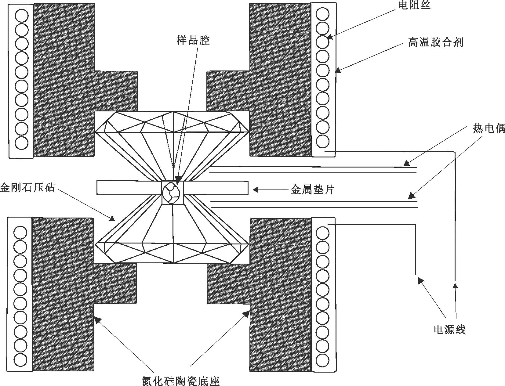
3.3高压容器1984年Roedder首次提出可以运用热液金刚石压腔(Hydrothermal Diamond-anvil Cell, HDAC)对熔体包裹体进行加热实验的可能。此后随着高温高压实验设备的更新迭代,通过HDAC装置对熔体包裹体进行加热,已成为目前唯一可以在均一实验期间可以原位观察到高内压熔体包裹体相变的方法(Li et al., 2014)。另外,与其他传统方法相比,使用HDAC设备还可以通过提高外部压力防止高内压熔体包裹体在加热过程中发生爆裂(Student et al., 2004)。目前使用的V型HDAC原理图如图3,上下两颗砧面平行的金刚石压合在存在孔洞的金属垫片上,形成密闭的样品腔,样品腔可装载1 mm×1 mm的样品,其深度由于金刚石对垫片的压合作用而小于0.25 mm(王新彦等, 2015)。因此,实验中需选择厚度小于0.25 mm的熔体包裹体,其样品腔和观察窗之间的距离比早期类型的更短,其距离仅为12 mm,有可能通过工作距离较长的物镜连续观察加热和熔体包裹体均一化过程(李胜虎等, 2015)。另外,使用HDAC进行均一化实验需要仔细遵循许多步骤,包括样品腔准备、样品制备、样品装载、压力调整、预热和正式加热等(王新彦等, 2015)。李建康等(2014)提出对甲基卡伟晶岩矿床花岗岩样品中的熔体包裹体均一化实验最好使用基于HDAC的方法进行,因为外部压力适当升高和较短的加热时间有助于抑制熔体包裹体发生成分泄漏,并且允许在均一化试验期间进行实时观察。此外,使用HDAC方法还有其他优势:可以精确控制加热速率,防止样品氧化;可以通过激光拉曼同步辐射X射线等原位微束分析技术对样品进行定性或定量分析(李胜虎, 2021)。例如,有学者应用金刚石热液压腔还原流体出溶的高温高压环境,配合原位拉曼光谱技术,揭示出岩浆-热液过渡阶段钨的主要赋存方式,并在模拟的原始温度压力条件下,获得钨在流体和熔体间的分配系数,进而探讨了钨在流体出溶过程中富集规律(Qiu et al., 2022)。
图3 V型HDAC设备原理图(据Li et al., 2014修改)
Fig. 3 Schematic diagram of the type-V HDAC equipment (modified after Li et al., 2014)
总之,基于HDAC的方法可以克服Linkam高温热台只能在大气压下进行加热实验的缺点,而未来随着HDAC设备的淬火方法和温度控制的发展,将进一步提高该方法在熔体包裹体均一实验中的应用(Li et al., 2014)。
3.4熔体包裹体均一化分析需要注意的问题通过系统梳理已有文献发现,虽然学者们对熔体包裹体进行加热实验时所使用的技术各有不同,但是研究结果显示不同技术所获取的熔体包裹体物化条件并没有显著差异,表明不同研究方法都能较好地实现熔体包裹体测温研究(Fedele et al., 2003; Student et al., 2004; Nielsen, 2011)。然而,在处理花岗岩样品时,避免使用淬火炉或高温容器,这是由于高温下熔体包裹体和主矿物可能会发生部分熔融,导致熔体包裹体甚至整个主矿物无法继续完成测试。
建议应用安装在显微镜上的高温台对熔体包裹体进行加热实验,可以在整个过程中连续观察包裹体瞬息的相态变化,能够更精确地获取均一温度。但是,对于玄武岩中在超高压下形成的熔体包裹体,一个大气压条件下的实验方法可能无法达到均一化,这种情况优选高压容器进行加热实验,以避免内外压力差过高导致包裹体发生爆裂(Cannatelli et al., 2016)。
4熔体包裹体成分测试随着分析技术的不断革新,人们逐渐掌握了熔体包裹体的成分分析技术,拓宽了在熔体包裹体中可获取信息的范围。熔体包裹体成分分析的取样方式与流体包裹体相同,可分为包裹体群分析和单个包裹体原位分析。对包裹体群进行整体分析较为简单快速,但是因为包裹体群可能由不同世代的包裹体共同组成,所获得数据为混合信息,无法反映单一期次的包裹体信息(孙贺等, 2009)。而选择不同世代的单个包裹体分别进行测试所得到的数据更加精细,可以避免群体成分分析法出现的成分混杂问题,是常用的也是前沿的包裹体研究手段(付乐兵等, 2015)。
单个包裹体激光原位分析最早始于20世纪80年代初,主要用于检测包裹体内金属元素的含量。随着分析技术手段的进步,不断推动熔体包裹体研究的发展。目前开展熔体包裹体成分分析的方法有扫描电子显微镜(Scanning Electron Microscope, SEM)、电子探针(Electron Probe Microanalysis, EPMA)、二次离子探针(Secondary Ion Mass Spetrometry, SIMS)、激光剥蚀电感耦合等离子体质谱仪(Laser Ablation Inductively Coupled Plasma Mass Spectrometer, LA-ICP-MS)、显微激光拉曼光谱(Micro-Raman Spectra, MRS)等,其中EPMA、SIMS和LA-ICP-MS可以对玻璃质(包括实验加热均一化后的样品)和已发生结晶的熔体包裹体进行主量元素、微量元素、挥发分或同位素的分析。Mason等(2008)通过对来自同一橄榄石内的一组成分相同(或近似)的熔体包裹体分别利用EPMA、SIMS和LA-ICP-MS(局部分析)对其成分分析,结果显示除部分微量元素(Pb、Th、Nb和U)外,3种方法获得的主、微量元素组成接近一致,证明了熔体包裹体成分分析技术的可靠性。下文将简要概述这几种方法的特点和应用。
图4 含有丰富流体内含物的明亮发光区的复合图像(据Baline et al., 2007)
Fig. 4 Composite image of a bright luminescent zone containing abundant inclusions (after Baline et al., 2007)
4.1扫描电子显微镜(SEM)扫描电子显微镜(SEM)是地质学领域常用的实验手段之一,可用于矿物相的识别和鉴定,在熔体包裹体的研究中也是非常有效的工具。扫描电镜可集成背散射电子探头(BSE)、X射线能谱(EDS)、阴极发光荧光谱仪(CL)、背散射电子衍射仪(EBSD)等附件(陈莉等, 2015)。其中,使用背散射电子(BSE)的成像是岩石学中最常用的方法,该技术可用于下一步成分分析之前检查待测包裹体的均一性,并使用能量色散X射线光谱(EDS)或波长色散X射线光谱(WDS)创建包裹体的元素分布图(Klébesz et al., 2012; Chen Y et al., 2011; Chen W et al., 2013)。但是,使用扫描电子显微镜(SEM)时必须注意因为元素在被电子束照射时可能会发生位移而产生误差:例如F、Cl、Ca和P在磷灰石和玻璃中变得活化;Na和K在斜长石和玻璃中变得活化(Cannatelli et al., 2016)。另外,扫描电镜成像使用阴极发光荧光谱仪(CL)可以捕获由电子束与样品表面相互作用产生的光子,例如Marie(2007)从格拉斯伯格斑岩矿床中采集的热液石英样品图像(图4),其中,明亮的CL区是由晶体生长期间捕获的大量流体包裹物生成的。这为研究石英中的熔体包裹体,提供了新的思路和可能,尤其是更精准地识别熔体包裹体。最近,国内学者将扫描电镜与共焦距拉曼成像联用对熔体包裹体进行逐层扫描,重建了熔体包裹体三维空间分布,成功获取熔体包裹体内部不同相态的体积分数,可为推断熔体包裹体捕获温度和岩浆房深度提供依据(Yuan et al., 2022)。
4.2电子探针分析(EPMA)电子探针(EPMA)的工作原理是将电子束聚焦于样品表面,激发样品组成元素产生特征性X射线,然后将样品X射线的波长和强度与标样对比,从而获得对样品的定量分析结果(Blundy et al., 2008)。该方法主要用于研究物质表面的元素组成及分布,是研究地球物质组成最基础的微束分析技术,也是最早应用于熔体包裹体成分研究的分析方法(张迪等, 2019;陈意等, 2021)。例如Carron(1961)通过电子探针尝试测定了熔体包裹体中的主量成分(Xia et al., 1991)。其工作原理是将电子束聚焦于样品表面,激发样品组成元素产生特征性X射线,然后将样品X射线的波长和强度与标样对比,从而获得样品的定量分析结果。EPMA可以分析熔体包裹体主量和部分微量元素以及挥发分组成(例如Si、Ti、Al、Fe、Mg、Mn、Ca、Na、K、Cr、P、Cl、F和S)(Kent, 2008),同时可以通过“差值法”计算H2O含量,为进一步限定包裹体最小捕获压力提供前提条件(张道涵等, 2017;李菊景等, 2022)。
EPMA的优势在于:①测试方法简单,且大多为无损测试,可对同一包裹体进行多次实验;②空间分辨率较高,可以斑束小至1~2μm,其中主量元素分析精度优于5%,而对于微量元素分析精度略差(10%~50%),具体取决于元素含量(Audétat et al., 2014)。该分析方法的局限在于只能对暴露至表面、内部均一的熔体包裹体成分分析。由于仪器硬件设备的发展和测试软件的更新,电子探针已不再满足于仅测试样品的主量元素,开始追求“微区、微量、高精度”的测试目标,以支撑地学前沿领域研究需求(张迪等, 2019)。
目前已有大量应用电子探针进行熔体包裹体研究的报道,徐九华等(2012)对乌拉嘎金矿含金斑岩体的石英斑晶中的熔体包裹体电子探针分析,表明SiO2含量较高,属酸性岩浆熔体;楚亚婷等(2016)对矿化斑岩中不同期次的玻璃质熔体包裹体展开电子探针测试,显示富碱高钾贫硅含铝的特征,并推断含矿地幔流体性质变化与熔流体不混溶作用有关;Jian等(2021)选取小秦岭脉状金矿区内含金石英矿脉中的富Au-Ag-Te熔体包裹体,通过包裹体测温、扫描电镜和电子探针分析手段,发现富碲熔体包裹体中的金属液滴能够从流体中汲取金进而导致富集成矿。相关研究显示了电子探针分析方法在熔体包裹体研究中具有重要的作用。
4.3二次离子探针(SIMS)20世纪80~90年代,地质学家开始使用离子探针(SIMS)对熔体包裹体展开成分分析。在真空下,使用直径为10~30 μm的一次离子束(如O-或Cs+)溅射包裹体样品表面,利用质谱仪收集和分析二次离子,最后通过法拉第杯或电子倍增器收集和测量经过质谱分离的二次离子强度,将其与标样对比,进而获得样品的元素、同位素丰度和比值等信息(Kent, 2008; Audétat et al., 2014)。SIMS具有较高的空间分辨率(10~30 μm),除了可以测定主、微量元素,还能够测量挥发性元素(H、C、F、S、Cl)、稳定同位素(H、Li、B、O、C、S、Cl)特征,并且是第一个用来分析熔体包裹体Pb同位素组成的分析技术(Zhang et al., 2020)。对于含量>(1~10)×10-6的元素,其分析精度一般优于5%~10%,而对于含量更低的元素,分析精度为10%~40%(Kent, 2008)。另外该方法对样品的破坏性较小,剥蚀深度在2~5 μm,可以在测试后保留样品做下一步分析(Cannatelli et al., 2016)。相对而言,其缺点在于:①样品制备较为繁琐,需要将包裹体抛光至表面;②分析效率较低,每次测试时间在30~90 min;③实验结果受基体效应影响较大。另外,在运用SIMS对包裹体进行成分分析时,内部标准元素的选择非常关键,通常使用电子探针测量熔体包裹体中的Si元素浓度作为其内标。
目前国外研究人员已经采用SIMS开展了大量熔体包裹体微量元素含量等研究。例如,有学者利用玄武岩中橄榄石和斜长石斑晶中的熔体包裹体示踪岩浆的起源与演化(Manfredo Capriolo et al., 2020)。Cooper等(2020)利用CAMECA IMS-1270型SIMS测试了岛弧火山岩单斜辉石中熔体包裹体的挥发分、微量元素及B同位素组成,探讨了深部熔体来源。相比之下,目前国内对于该方面的研究报道较少。张乐等(2019)应用中国科学院广州地球化学研究所的CAMECA IMS 1280-HR型SIMS对夏威夷玄武岩中橄榄石捕获的熔体包裹体进行了分析测试,结果与全岩数据显示了较好的一致性,证明了CAMECA IMS 1280-HR型SIMS可以作为分析熔体包裹体微量元素的有效工具,并建立了相应的微量元素数据处理包SIMSTraElement,可以对SIMS测试的微量元素数据进行快速便捷处理。
4.4激光剥蚀电感耦合等离子体质谱仪(LA-ICP-MS)使用激光剥蚀电感耦合等离子体质谱仪方法(LA-ICP-MS)对单个包裹体原位分析的方法已有40余年的研究历史(Bennet et al., 1980; Deloule et al., 1982; Tsui et al., 1979)。最早使用激光剥蚀系统与摄谱仪联用分析剥蚀产物的光谱特征实现元素的定性-半定量分析,随后与电感耦合等离子质谱仪(ICP-MS)和原子发射光谱仪(ICP-AES)等联用对单个包裹体展开定量分析(Günther et al., 1998)。随着激光剥蚀技术的发展,在21世纪初期,学者们尝试通过激光剥蚀电感耦合等离子体质谱仪(LA-ICP-MS)对多相且未暴露至表面的单个熔体包裹体进行成分分析,并全面总结了基本原则和数据分析方法(Halter et al., 2002; Pettke, 2006; Mason et al., 2008)。目前,LA-ICP-MS已成为分析单个熔体包裹体成分的前沿测试方法(Zhang et al., 2020)。值得一提的是,南京大学内生金属矿床成矿机制研究国家重点实验室建立了单个熔体包裹体LA-ICP-MS微区分析方法,可以自主完成单个熔体包裹体成分数据的测试,具备国际先进水平的单个包裹体成分分析技术(倪培等, 2021)
LA-ICP-MS的工作原理为利用脉冲激光束对样品表面进行剥蚀,使之熔蚀气化,形成气溶胶,同时输入惰性气体(He或Ar)作为载气将剥蚀物质传送至电感耦合等离子体中快速离子化,再经过质谱仪,根据质荷比进行过滤,最后利用接收器的检测系统对不同质荷比的离子信号强度进行分析,如此可获得样品成分的瞬时信号。LA-ICP-MS对于分析样品微量元素具有较大的优势,元素周期表中除F、O、N、H及惰性气体外的其他元素都可以用LA-ICP-MS进行分析,但针对不同元素的检测限差异很大(Kent, 2008; Pettke et al., 2012)。考虑到激光斑束的特点,在选择待测包裹体时要尽量选择形状接近于球形的包裹体,最佳包裹体尺寸为20~50 µm(郭伟等, 2020),并且距主矿物表面适当浅深度,以便激光可以将其完整剥蚀从而获得更准确的结果。另外,样品切片的厚度尽量控制在100~200 µm,避免包裹体片太薄导致激光打穿而破坏剥蚀池。
目前应用LA-ICP-MS对单个熔体包裹体进行成分分析的方法分为局部剥蚀和整体剥蚀2种(孙贺, 2014)。局部分析法与EPMA和SIMS类似,主要针对直径较大、内部成分均一且暴露于表面的熔体包裹体,测试过程中激光直接剥蚀到熔体包裹体,因此分析精度较高(优于5%~10%)。另一种包裹体整体剥蚀的方法在分析结晶质熔体包裹体时无需事先均一化,其工作原理是在测试过程中,激光首先照射在主矿物上,再穿透主矿物对其内部熔体包裹体进行剥蚀(图5)。所以该方法所得到的信号是部分主矿物+熔体包裹体的成分,因此需要通过选择合理内标测得主矿物在混合信号中的质量比例,在数据处理时将主矿物的贡献量扣除,才能得到更准确的包裹体成分信息,这种方法的优点是可容许的剥蚀深度大(可达100 µm),不需要提前均一熔体包裹体,避免了均一过程中发生的物质泄露和损耗;缺点是该方法计算时容易引入误差,所得数据的精度和准确度相比局部分析法较差(Pettke et al., 2012)。
近年来,国内学者应用LA-ICP-MS分析单个熔体包裹体元素组成的报道与日俱增,孙艺等(2011)通过LA-ICP-MS对不同岩相带内石英中的熔体包裹体进行原位研究,证实了岩浆残余富集和流体叠加对稀土元素配分的改造作用;Zhang等(2017)对美国Bingham Canyon斑岩型Cu-Mo-Au矿床中的单个熔体包裹体进行原位分析,重建了成矿岩浆中金属和硫的含量,并得出成矿岩浆中并不富含Cu的结论;Chang等(2020)运用LA-ICP-MS对不同侵入体和不同寄主矿物中的熔体包裹体进行整体剥蚀分析,并对所得数据开展系统评估,结果表明相比其他分析方法,LA-ICP-MS是目前获得中基性深成侵入体中熔体包裹体内未改造元素含量最直接有效的手段;刘晓阳(2022)对胶东地区与金成矿有关的中基性岩脉中的硫化物熔体包裹体展开LA-ICP-MS原位微量元素成分测试,结果表明岩浆源区壳幔混合过程中的物理化学变化导致硫化物熔体包裹体出熔,并对后期成矿起到了金属预富集作用。
4.5显微拉曼光谱(MRS)20世纪70年代中期,科研人员开始尝试使用激光拉曼光谱(MRS)对流体包裹体展开测试(Delhaye et al., 1975)。随着几十年来地质学家们的不断探索,显微激光拉曼已成为熔体包裹体化学分析的一种基本方法,通常用于测量熔体包裹体气泡中的挥发分含量及子矿物相成分(Bodnar et al., 2020)。其工作原理是当激光穿过待测样品时,部分散射光频率发生改变,这种现象称为“拉曼散射”,其中改变了频率的光谱称为拉曼光谱。通常将光学显微镜与拉曼光谱仪联用,使用高倍物镜聚焦于包裹体样品,对包裹体中的不同相态部分进行定性和半定量分析(陈勇等, 2006)。
拉曼光谱具有空间分辨率高(1~2 μm)、低成本、无损且原位(与激光辐射反应的样品除外)、制样简单、不需要将包裹体抛光至表面的优点(Chou et al., 2017),并且适用于花岗岩中的含水量较高的包裹体,其最优条件下测H2O的准确度为± 0.1%(质量分数);其缺点在于,荧光效应较强,计算过程较为复杂,需要设定与待测样品成分相近的标样(Muro et al., 2006; Severs et al., 2007)。在应用拉曼光谱测定挥发分含量时需要注意,如果熔体包裹体中气泡的密度较低,那么用其测定挥发分含量时要将包裹体和气泡到样品表面的距离控制在30 μm内,以避免由于包裹体位置过深导致的拉曼信号弱,所得数据不佳(Cannatelli et al., 2016)。
本世纪以来,中国学者通过拉曼光谱技术对熔体包裹体进行了大量研究,Xie等(2009)对牦牛坪稀土矿内石英和萤石中的熔体包裹体及共存的流体包裹体中的CO2、液相和子晶成分进行了研究,结合测温数据探讨了成矿流体的起源和演化过程。另外,有学者分别对中国北方和长江中下游地区多个代表性矽卡岩型矿床中的熔体包裹体进行了激光拉曼测试,结果表明包裹体内固相成分与寄主矿物一致,认为该类矿化矽卡岩为岩浆成因,矽卡岩岩浆为成矿提供了物质来源(赵劲松等, 2015;赵斌等, 2017);赵岩岩等(2020)在湖北铜绿山铜铁(金)矿床矽卡岩带的熔融包裹体中检测到石榴子石、方解石、赤铁矿和水,推断早期流体已经富集了足够的铁。近期,有研究者通过拉曼光谱测试在橄榄石内富CO2的熔体包裹体中发现了菱镁矿子矿物,认为其是寄主矿物橄榄石在特定温度下与富CO2流体反应而沉淀形成的次生矿物,论证了岩石圈地幔吸收了大量的CO2,是碳循环的“碳陷阱”(Xu et al., 2023)。另外,有国外学者首次提出将拉曼3D成像新技术与X射线荧光光谱仪和扫描电子显微镜联合应用,成功识别和测量出玻璃质熔体包裹体中气泡的成分分布特征,发现气泡内壁上的细小结晶相,这项研究表面对熔体包裹体中的收缩气泡进行三维识别有利于更准确评估岩浆中的喷发前的挥发分含量(Schiavi et al., 2020)。
图5 单个熔体包裹体(MI)的LA-ICP-MS分析示意图(据Pettke et al., 2006修改)
Fig. 5 Sketch map showing the LA-ICP-MS analysis of a single melt inclusion (MI) (modified from Pettke et al., 2006)
4.6熔体包裹体成分分析需要注意的问题(1)做微区分析前要提前熟知相关方法,不同方法对于所测包裹体的选择要求也不同,研究人员应明确自己的实验需求,确定测试前包裹体样品的切片方式、仔细筛选包裹体组合或单个包裹体以及是否需要将包裹体加热至均一化等工作。
(2)所有的微束技术都是相对的分析方法,对于EPMA、SIMS等仪器,检测根据已知元素丰度作为标准进行校准是实验顺利进行的先决条件,因此针对不同的分析技术和实验对象,选择合适的校正方法尤为关键。并且测试流程中涉及信号强度(束斑大小、频率、强度)、分馏效应、基体效应等一系列信息干扰,研究人员要综合考虑各种情况,对相应干扰采取预防措施。
(3)使用微区成分分析所得结果的不确定度和准确性可能取决于许多因素,包括分析的元素、光束、分析条件和软件及人为误差等。例如,由于铜在硅酸盐熔体中的扩散速度很快,如果寄主矿物外界岩浆的铜含量发生了变化,则熔体包裹体将会和岩浆发生扩散再平衡导致熔体包裹体内部铜含量偏离初始值(Zhang et al., 2023)。因此在数据处理要格外注意。建议实验人员将所得包裹体数据与全岩化学数据相对比,以保证实验数据是可靠的。
5总结和展望国际上关于熔体包裹体的分析已走过几十年的研究历程,但中国的研究尚处于起步阶段,尤其受制于分析技术和分析方法的薄弱。然而,熔体包裹体保存有原始的岩浆成分信息,并记录了各瞬时的岩浆演化信息,在岩石成因研究中独具优势,加强其研究对解决地球科学问题迫在眉睫。因此,系统梳理熔体包裹体的分析技术势在必行。
综合国内外研究进展,针对熔体包裹体的系统分析总体可以分为3个步骤:①在光学显微镜下对熔体包裹体进行岩相学观察,尽量选取大小适合(>10μm)、形态近似球形、并且符合研究内容所需的熔体包裹体进行下一步测试;②熔体包裹体显微测温,通过一个大气压下的管式淬火炉、高温热台、高压容器等对包裹体进行加热实验使其内部均一化,在获取成岩成矿流体的温度同时,便于展开其他实验研究;③熔体包裹体成分分析,利用SEM、EPMA、SIMS或LA-ICP-MS对包裹体内部组成进行仔细筛查和甄别。
随着获取单个熔体包裹体的主、微量成分的技术设备和方法不断完备,该方法相比于全岩地球化学更为丰富的岩浆成分信息,揭示出传统岩石地化手段难以识别的地质过程信息,正日益成为探讨岩浆起源和演化不可或缺的重要手段。尤其是精确测定熔体包裹体中同位素组成的工作方兴未艾,目前已成功应用SIMS和LA-ICP-MS开展了熔体包裹体中H、Li、B、C、O、S、Cl、Sr、Nd、Pb同位素研究,在揭示地壳深循环、地幔源区不均一性、地壳岩浆混染过程等方面显示出巨大潜力。另外,熔体包裹体是揭示岩浆挥发分含量(如H2O、CO2、F、S、Cl等)至关重要的手段,在研究火山活动及其环境效应方面正发挥着不可替代的作用,为深入认识全球碳-水循环、气候变化和生物灭绝事件等重大关切问题提供了新的视角。尤其值得注意的是,熔体包裹体可以提供珍贵的原始岩浆中成矿元素和重要络合物含量信息,是研究成矿过程的有力手段。
致 谢感谢主编及匿名审稿专家对本文提出的宝贵意见和建议,使得文章内容得以提升。
-
参考文献
Anderson A T. 1991. Hourglass inclusions; theory and application to the Bishop rhyolitic tuff[J]. American Mineralogist, 76(3-4): 530-547.
Audétat A and Lowenstern J B. 2014. Melt inclusions[D]. In: Turekian, H D, Holland K K. (Eds.), Treatise on Geochemistry, 2nd ed. Elsevier, Oxford. 143-173.
Baline and Marie L. 2007. Hydrothermal fluids and Cu-Au mineralization of the deep grasberg porphyry deposit, Papua, Indonesia[D]. Texas: The University of Texas. 1-268.
Bennett J N and Grant J N. 1980. Analysis of fluid inclusions using a pulsed laser microprobe[J]. Mineralogical Magazine, 43(331): 945-947.
Blundy J and Cashman K. 2008. Petrologic reconstruction of magmatic system variables and processes[J]. Reviews in Mineralogy &Geochemistry, 69(1): 179-239.
Bodnar R J and Frezzotti M L. 2020. Microscale chemistry: Raman analysis of fluid and melt inclusions[J]. Elements, 16(2): 93-98.
Bodnar R J, Student J J and Webster J D. 2006. Melt inclusions in plutonic rocks: Petrography and microthermometry[J]. Melt inclusions in plutonic rocks, 36: 1-25.
Cannatelli C, Lima A, Bodnar R J, De Vivo B, Webster J D and Fedele L. 2007. Geochemistry of melt inclusions from the Fondo Riccio and Minopoli 1 eruptions at Campi Flegrei (Italy)[J]. Chemical Geology, 237(3): 418-432.
Cannatelli C. 2012. Understanding magma evolution at Campi Flegrei (Campania, Italy) volcanic complex using melt inclusions and phase equilibria[J]. Mineralogy and Petrology, 104: 29-42.
Cannatelli C, Doherty A L, Esposito R, Lima A and Vivo B D. 2016. Understanding a volcano through a droplet: A melt inclusion approach[J]. Journal of Geochemical Exploration, 4-19.
Capriolo M, Marzoli A, Aradi L E, Callegaro S, Dal Corso J, Newton R J, Mills B J W, Wignall P B, Bartoli O, Baker D R, Youbi N, Remusat L, Spiess R and Szabó C. 2020. Deep CO2 in the End-Triassic central atlantic magmatic province[J]. Nature Communications, 1-11.
Carron J P. 1961. Premieres donnees sur la composition de certains reliquats magmatiques[J]. Comptes Rendus de l'Académie des Sciences, 53: 2016-2018.
Chang H L and Huang H L. 1998. Characteristics and origin of melt-fluid inclusion[J]. Bulletin of Geological Science and Technology, 17(S1):36-39(in Chinese with English abstract).
Chang J and Audétat A. 2020. LA-ICP-MS analysis of crystallized melt inclusions in olivine, plagioclase, apatite and pyroxene: Quantification strategies and effects of post-entrapment modifications[J]. Journal of Petrology, 62(4): 1-23.
Chen L, Xu J and Chen J. 2015. Applications of scanning electron microscopy in earth sciences[J]. Science China: Earth Sciences, 58: 1768-1778(in Chinese with English abstract).
Chen W, Kamenetsky V S and Simonetti A. 2013. Evidence for the alkaline nature of parental carbonatite melts at Oka complex in Canada[J]. Nature Communication, 4: 2687.
Chen Y, Zhao Y Q, Yan S S, Liu Y C and Wang Q. 2006. The application of laser raman spectroscopy to obtaining internal pressure of fluid inclusions[J]. Acta Geoscientica Sinica, 27(1): 69-73(in Chinese with English abstract).
Chen Y, Provost A, Schiano P and Cluzel N. 2011. The rate of water loss from olivinehosted melt inclusions[J]. Contributions to Minera-logy and Petrology, 162: 625-636.
Chen Y, Hu Z C, Jia L H, Li J H, Li Q L, Li X G, Li Z P, Long T, Tang X, Wang J, Xia X P, Yang W, Yuan J Y, Zhang D and Li X H. 2021. Progress of microbeam analytical technologies in the past decade (2011-2020) and prospecrt[J]. Bulletin of Mineralogy,Petrology and Geochemistry, 40(1): 1-35(in Chinese with English abstract).
Chou I M and Wang A. 2017. Application of laser Raman micro-analyses to earth and planetary materials[J]. Journal of Asian Earth Sciences, 145: 309-333.
Chu Y T. 2016. Evolution of fluids in deep processes: Petrogenesis and metallogensis of Cenozoic alkaline-rich magma in western Yunnan Province[D]. Chengdu: Chengdu University of Technology. 1-216(in Chinese with English abstract).
Clocchiatti R. 1975. Les inclusions vitreuses des cristaux de quartz. Etude optique, thermo-optique et chimique[J]. Applications Géologiques, 122: 1-96(in French).
Cooper G F, Macpherson C G, Blundy J D, Maunder B, Allen R W, Goes S, Collier J S, Bie L, Harmon N, Hicks S P, Iveson A A, Prytulak J, Rietbrock A, Rychert C A, Davidson J P and the VoiLA team. 2020. Variable water inputcontrols evolution of the Lesser Antilles volcanic arc[J]. Nature, 582(7813): 525-529.
Danyushevsky L V, McNeill A W and Sobolev A V. 2002. Experimental and petrological studies of melt inclusions in phenocrysts from mantle-derived magmas: An overview of techniques, advantages and complications[J]. Chemical Geology, 183(1-4): 5-24.
Davidson P and Crawford A J. 2006. Coexisting melt and fluid inclusions at Batu Hijau: Evidence for primary magmatic aqueous fluids and their relationship to mineralisation[J]. Geochimica et Cosmochimica Acta, 70(18-supp-S): A131-A131.
Delhaye M and Dhamelincourt P. 1975. Raman microprobe and microscope with laser excitation[J]. Raman Spectrosc, 3: 33-43.
Deloule E and Eloy J F. 1982. Improvements of laser probe mass spectrometry for the chemical analysis of fluid inclusions in ores[J]. Chemical Geology, 37(1-2): 191-202.
Esposito R, Klebesz R and Bartoli O. 2012. Application of the Linkam TS1400XY heating stage to melt inclusion studies[J]. Central European Journal of Geosciences, 4(2):208-218.
Esposito R, Hunter J, Schiffbauer J D and Bodnar R J. 2014. An assessment of the reliability of melt inclusions as recorders of the pre-eruptive volatile content of magmas[J]. American Mineralogist, 99(5-6): 976-998.
Fedele L, Bodnar R J, De Vivo B and Tracy R J. 2003. Melt inclusion geochemistry and computer modeling of trachyte petrogenesis at Ponza, Italy[J]. Chemical Geology, 194: 81-104.
Fu L B, Wei J H, Zhang D H, Tan J, Tian N and Zhao Z X. 2015. A review of LA-ICPMS analysis for individual fluid inclusions and its applications in ore deposits[J]. Journal of Central South University(Science and Technology), 46(10): 3832-3840(in Chinese with English abstract).
Günther D, Audétat A, Frischknecht R and Heinrich C A. 1998. Quantitative analysis of major, minor and trace elements in fluid inclusions using laser ablation inductively coupled plasma mass spectrometry[J]. Journal of Analytical Atomic Spectrometry, 13(4): 263-270.
Guo W, Lin X and Hu S H. 2020. Advances in LA⁃ICP⁃MS Analysis for individual fluid inclusions and applications [J]. Earth Science, 45(4): 1362-1374(in Chinese with English abstract).
Halter W E, Pettke T and Heinrich CA. 2002. The origin of Cu/Au ratios in porphyry-type ore deposits[J]. Science, 296(5574): 1844-1846.
Hamada M, Iwamori H, Brandl P A, Ushikubo T, Shimizu K, Ito M, Li H and Savov I P. 2020. Temporal evolution of proto-Izu-BoninMariana arc volcanism over 10 Myr: Constraints from statistical analysis of melt inclusion compositions[J]. Journal of Petrology, 61: 22-109.
Humphreys M C S, Menand T, Blundy J D and Klimm K. 2008. Magma ascent rates in explosive eruptions: Constraints from H2O diffusion in melt inclusions[J]. Earth and Planetary Science Letters, 270(1-2): 25-40.
Jian W, Mao J W, Lehmann B, Cook N J, Xie G Q, Liu P, Duan C, Alles J and Niu Z J. 2021. Au-Ag-Te-rich melt inclusions in hydrothermal gold-quartz veins, Xiaoqinling lode gold distric, central China[J]. Econ. Geol., 116(5): 1239-1248.
Johnson M C, Anderson A T and Rutherford M J. 1994. Pre-eruptive volatile contents of magmas[J]. Reviews In Mineralogy & Geochemistry, 30(1): 281-330.
Kent A J R. 2008. Melt inclusions in basaltic and related volcanic rocks[J]. Reviews In Mineralogy & Geochemistry, 69(1): 273-331.
Klébesz R, Bodnar R, De Vivo B, Török K, Lima A and Petrosino P. 2012. Composition and origin of nodules from the≈20 ka Pomici di Base (PB)-Sarno eruption of Mt. Somma-Vesuvius, Italy[J]. Central European Journal of Geosciences, 4(2): 324-337.
Kyle J R and Ketcham R A. 2015. Application of high resolution X-ray computed tomography to mineral deposit origin, evaluation, and processing[J]. Ore Geology Reviews, 65(4): 821-839.
Li J J, Guo Z F, Zhao W B and Zhang M L. 2022. Application of melt inclusions for studying volcanic CO2 fluxes[J]. Bulletin of Mine-ralogy, Petrology and Geochemistry, 41(4): 711-728(in Chinese with English abstract).
Li J K and Li S H. 2014. Application of hydrothermal diamond anvil cell to homogenization experiments of silicate melt inclusions[J]. Acta Geologica Sinica-English Edition, 88(3): 854-864.
Li J K, Zhang D H and Li S H. 2011. Application of melt inclusions to estimating ore-forming pressure (depth) of granite-related ore deposits[J]. Mineral Deposits, 30(6): 1002-1016(in Chinese with English abstract).
Li S H, Xiong Y X, Shan W, Shu L and Chi N J. 2021. Brief introduction of high temperature and high pressure experimental platform for HDAC[J]. Shandong Land and Resources, 37(1): 50-52(in Chinese with English abstract).
Li S H, Li J K and Zhang D H. 2015. Application of hydrothermal diamond-anvil cell in fluid inclusions: An example from the Jiajika pegmatite deposit in western Sichuan, China[J]. Acta Geologica Sinica, 89(4): 747-754(in Chinese with English abstract).
Liu X Y. 2023. Deep magmatic process of the Early Cretaceous gold metallogenic system in the eastern North China Craton: Evidence from in-situ geochemical analyses of minerals and melt inclusions from mantle xenoliths and intermediate-basic dykes[D]. Wuhan:China University of Geosciences. 1-129(in Chinese with English abstract).
Liu Y, Anderson A T and Wilson C J N. 2007. Melt pockets in phenocrysts and decompression rates of silicic magmas before fragmentation[J]. Journal of Geophysical Research: Solid Earth, 112(B6), B06204.
Lowenstern J B. 1995. Applications of silicate melt inclusions to the study of magmatic volatiles[A]. In: Thompson J F H, ed. Magmas, Fluid and Ore Deposits vol. 23. Mineralogical Society of Canada Short Courses[C]. 71-99.
Lowenstern J B. 1994. Dissolved volatile concentrations in an ore-forming magma[J]. Geology, 22: 893-896.
Marie B L. 2007. Hydrothermal fluids and Cu-Au mineralization of the Deep Grasberg porphyry deposit, Papua, Indonesia[D]. Texas: University of Texas at Austin. 1-268.
Mason P R D, Nikogosian I K and Bergen M J V. 2008. Major and trace element analysis of melt inclusions by laser ablation ICP-MS[J]. Laser Ablation ICP-MS in the Earth Sciences 40: 219-240.
Muro A D, Giordano D, Villemant B, Montagnac G, Scaillet B and Romano C. 2006. Influence of compo- sition and thermal history of volcanic glasses on water content as de-termined by micro-Raman spectrometry[J]. Applied Geochemistry, 21(5): 802-812.
Ni P, Fan H R, Pan J Y, Chi Z and Cui J M. 2021. Progress and prospect of fluid inclusion research in the past decade in China (2011-2020)[J]. Bulletin of Mineralogy, Petrology and Geochemistry, 40(4): 802-818(in Chinese with English abstract).
Nielsen R L. 2011. The effects of re-homogenization on plagioclase hosted melt inclusions[J]. Geochemistry Geophysics Geosystems, 12(10): 1-16.
Pettke T, Oberli F, A Audétat, Guillong M, Simon A C, Hanley J J and Klemm L M. 2012. Recent developments in element concentration and isotope ratio analysis of individual fluid inclusions by laser ablation single and multiple collector ICP-MS[J]. Ore Geology Reviews, 44:8-10
Pettke T. 2006. In situ laser-ablation ICPMS analysis of melt inclusions and prospects for constraining subduction zone magmatism[R]. Short Course. 51-80.
Qiu Y, Wang X L, Lu J J, Chou I M, Wan Y, Zhang R Q, Zhang W L and Sun R. 2022. In situ observations of tungsten speciation and partitioning behavior during fluid exsolution from granitic melt[J]. Science Bulletin, 67(22): 2358-2368.
Richard A, Morlot C, Créon L, Beaudoin N, Balistky V S, Pentelei S, Dyja-Person V, Giuliani G, Pignatelli I, Legros H, Sterpenich J and Pironon J. 2018. Advances in 3D imaging and volumetric reconstruction of fluid and melt inclusions by high resolution X-ray computed tomography[J]. Chemical Geology, 508: 2-11.
Roedder E and Weiblen P W. 1970. Lunar petrology of silicate melt inclusions, Apollo 11 rocks. Proceedings of the Apollo 11 Lunar Science Conference[J]. Geochimica et Cosmochimica Acta, Supplement I: 801-837.
Roedder E. 1979. Origin and significance of magmatic inclusions[J]. Bulletin de Mineralogie, 102(56): 487-510.
Roedder E. 1992. Fluid inclusion evidence for immiscibility in magmatic differentiation[J]. Geochimica et Cosmochimica Acta, 56 (1): 5-20.
Schiano P. 2003. Primitive mantle magmas recorded as silicate melt inclusions in igneous minerals[J]. Earth Sciences Reviews, 63(1-2): 121-144.
Schiavi F, Bolfan Casanova N, Buso R, Laumonier M, Laporte D, Medjoubi K, Venugopal S, Gómez-Ulla A, Cluzel N and Hardiagon M. 2020. Quantifying magmatic volatiles by Raman microtomography of glass inclusion-hosted bubbles[J]. Geochemical Perspectives Letters, 16: 17-24.
Severs M J, Azbej T, Thomas J B, Mandeville C W and Bodnar R J. 2007. Experimental determination of H2O loss from melt inclusions during laboratory heating: Evidence from Raman spectroscopy[J]. Chemical Geology, 237(3-4): 358-371.
Sobolev A V, Dmitriev L V, Baruskov V L, Nevsorov V N and Slutsky A B. 1980. The formation conditions of the high-magnesian olivines from the monomineralic fraction of Luna 24 regolith[J]. Proceedings of the Apollo 11 Lunar Science Conference, Geochimica et Cosmochimica Acta, SupplimentⅠ: 105-116.
Sobolev A V. 1996. Melt inclusions in minerals as a source of principle petrological information[J]. Petrology, 4(3): 209-220.
Sobolev V S and Kostyuk V P. 1975. Magmatic crystallization based on a study of melt inclusions[J]. Fluid Inclusion Research, 9: 182-253.
Sorby H C. 1858. On the microscopical, structure of crystals, indica-ting the origin of minerals and rocks[J]. Quarterly Journal of the Geological Society, 14(1-2): 453-500.
Sinton C W, Christie D M, Coombs V L, Nielsen R L and Fisk M R. 1993. Near-primary melt inclusions in anorthite phenocrysts from the Galapagos Platfrom[J]. Earth and Planetary Science letters, 119(4): 527-537.
Sun H and Xiao Y L. 2009. Fluid inclusion: Latest development, geological applications and prospect[J]. Advances in Earth Science, 24(10): 1105-1121(in Chinese with English abstract).
Sun H. 2014. Application of melt inclusions and Li isotope in earth sciences[D]. Heifei:University of Science and Technology of China.1-157(in Chinese with English abstract).
Sun Y, Lai Y, Chen J and Shu Q H. 2011. The impact of residual enrichment of magma and fluid stacking on rare earth partitioning:In Situ LA-ICP-MS study of molten inclusions[J]. Acta Mineralogica Sinica, 31(S1): 291-292(in Chinese with English abstract).
Student J J and Bodnar R J. 1999. Synthetic fluid inclusions XIV: Coexisting silicate melt and aqueous fluid inclusions in the haplogra-nite-H2O-NaCl-KCl system[J]. Journal of Petrology, 40(10): 1509-1525.
Student J J and Bodnar R J. 2004. Silicate melt inclusions in porphyry copper deposits: Identification and homogenization behavior[J]. The Canadian Mineralogist, 42(5): 1583-1599.
Tsui T F and Holland H D. 1979. The analysis of fluid inclusions by Laser Microprobe[J]. Econ. Geol., 74(7): 1647-1653.
Wallace P J, Plank T, Bodnar R J, Gaetani G A and Shea T. 2021. Olivine-hosted Melt inclusions: A microscopic perspective on a complex magmatic world[J]. Annual Review of Earth and Planetary Sciences, 49: 465-494.
Wang D, Lu H Z and Shan Q. 2017. Advances on melt inclusion studies[J]. Acta Petrologica Sinica, 33(2): 653-666(in Chinese with English abstract).
Wang G G, Ni P and Pan J Y. 2020. Fluid characteristics of granite-related ore forming systems[J]. Bulletin of Mineralogy, Petrology and Geochemistry, 39(3): 463-471(in Chinese with English abstract).
Wang X Y, Li J K, Li S H, Zhang D H and Bao B. 2015. A comparative study of applications of Linkam high temperature stage and Hydrothermal Diamond Anvil-Cell (HDAC) to homogenization experiment of melt inclusions[J]. Mineral Deposits, 34(3): 589-601(in Chinese with English abstract).
Wu Y M, Guo F, Wang X C, Wilde Simon A and Fan W M. 2020. Derivation of Jurassic HIMU-like intraplate basalts from mantle transition zone in South China: New geochemical constraints from oli-vine-hosted melt inclusion[J]. Lithos, 354: 105337.
Xia L Q, Liu W F and Tang N A. 1991. Abundances of F, CI, S and P in volcanic magmas and their evolution, Wudalianchi[J]. Chinese Journal of Geochemistry (English Language Edition), (2): 120-130.
Xie Y L, Hou Z Q, Yin S P, Simon C D, Xu J H, Tian S H and Xu W Y. 2009. Continuous carbonatitic melt-fluid evolution of a REE mi-neralization system: Evidence from inclusions in the Maoniuping REE deposit, western Sichuan, China[J]. Ore Geology Reviews, 36: 90-105.
Xu J H, Wei H, Wang Y H, Zeng Q D, Liu J M, Wang Y B and Mao Q. 2012. Sub-volcanic hydrothermal mineralization of the Wulaga gold deposit, Heilongjiang, China: Evidences from melt and fluid inclusions[J]. Acta Petrologica Sinica, 28(4): 1305-1316(in Chinese with English abstract).
Xu X, Tian W, Zhang L F and Li H J. 2023. Carbon enrichment in the lithospheric mantle: Evidence from the melt inclusions in mantle xenoliths from the Hainan basalts[J]. Acta Geologica Sinica (English), 97(1): 358-375.
Yuan J Y, Su W, Hu X M, Li X G and Fei C H. 2022. Application of Raman imaging and scanning electron microscopy techniques for the advanced characterization of geological samples[J]. Microscopy Research and Technique, 85(7): 2729-2739.
Yuan S D, Zhao P L and Liu M. 2020. Some problems involving in petrogenesis and metallogenesis of granite-related tin deposits[J]. Mineral Deposits, 39(4): 607-618(in Chinese with English abstract).
Zajacz Z and Halter W. 2007. LA-ICPMS analyses of silicate melt inclusions in coprecipitated minerals: Quantifification, data analysis and mineral/melt partitioning[J]. Geochimica et Cosmochimica Acta, 71(4): 1021-1040.
Zhang D, Chen Y, Mao Q, Su B, Jia L H and Guo S. 2019. Progress and challenge of electron probe microanalysis technique[J]. Acta Petrologica Sinica, 35(1): 261-274(in Chinese with English abstract).
Zhang D H and Audétat A. 2017. What caused the formation of the giant Bingham Canyon porphyry Cu-Mo-Au deposit? Insights from melt inclusions and magmatic sulfides[J]. Econ. Geol., 112(2): 221-244.
Zhang D H, Wei J H, Fu L B and Wang D Z. 2017. Formation, modification and analytical techniques of melt inclusion, and their application in Economic Geology[J]. Earth Science, 42(6): 990-1007(in Chinese with English abstract).
Zhang L, Qian S, Li N, Hong L, Zhang Y and Ren Z. 2020. Simultaneous in situ determination of Pb isotope ratios and trace element concentrations in melt inclusions by LA-ICP-MS[J]. Geochemistry, Geophysics, Geosystems, 22: 1-11.
Zhang L, Wang F Y and Ni H W. 2023. Anisotropic copper diffusion in olivine: Implications for Cu preservation in olivine and olivine-hosted melt inclusions[J]. Geochimica et Cosmochimica Acta, 347: 116-124.
Zhang L, Wang Q, Ding X and Li W C. 2021. Diverse serpentinization and associated abiotic methanogenesis within multiple types of olivine-hosted fluid inclusions in orogenic peridotite from northern Tibet[J]. Geochimica et Cosmochimica Acta, 296: 1-17.
Zhang L, Xia X P, Yang Q, Zhang W F, Xiong B Q, Qian S P and Ren Z Y. 2019. Determination of trace elements in melt inclusions by secondary ion mass spectrometry[J]. Geochimica, 48(1): 1-8(in Chinese with English abstract).
Zhao B, Zhao J S and Xu D R. 2017. Laser Raman analysis results of mineral inclusions from deposit of skarn type distributed in the Middle and Lower Reaches Metallogenic Belt of Yangtze River, China and their geological-geochemical significance[J]. Acta Petr-ologica Sinica, 33(6): 1841-1858(in Chinese with English abstract).
Zhao J S,Zhao B, Li J W, Xu D R, He M C, Zheng J P. 2015. Magma control of magmatic skarn to the formation of some deposits of skarn type: Evidence from Laser Raman analyses of inclusions in minerals[J]. Acta Petrologica Sinica, 31(4): 1079-1088(in Chinese with English abstract).
Zhao Y Y, Tan J, Liu X Y, Zhang M, Chen Y, He D. 2020. Inclusion features and geological significance of the Tonglüshan skarn-type copper-iron (gold) deposit in Daye, Hubei[J]. Bulletin of Geological and Technology, 39(6): 64-74(in Chinese with English abstract).
Zhu R X, Hou Z Q, Guo Z T and Wan B. 2021. Summary of “the past, present and future of the habitable Earth: Development strategy of Earth science”[J]. Chinese Science Bulletin, 66(35): 4485-4490(in Chinese with English abstract).
Zirkel F. 1873. Die mikroscopische beschaftenheit der mineralien und Gesteine[M]. Wilhelm Englemann, Leipzig(in German).
附中文参考文献
常海亮,黄惠兰. 1998.熔-流包裹体的特征及成因[J].地质科技情报, 17(S1):36-39.
陈莉,徐军,陈晶. 2015.扫描电子显微镜显微分析技术在地球科学中的应用[J].中国科学:地球科学, 45(9): 1347-1358.
陈意,胡兆初,贾丽辉,李金华,李秋立,李晓光,李展平,龙涛,唐旭,王建,夏小平,杨蔚,原江燕,张迪,李献华. 2021.微束分析测试技术十年(2011-2020)进展与展望[J].矿物岩石地球化学通报, 40(1): 1-35.
陈勇,周瑶琪,颜世永,刘英超,王强. 2006.激光拉曼光谱技术在获取流体包裹体内压中的应用及讨论[J].地球学报, 27(1): 69-73.
楚亚婷. 2016.深部地质过程中流体作用及流体演化研究——以滇西新生代富碱岩浆成岩成矿为例[D].成都:成都理工大学. 1-216.
付乐兵,魏俊浩,张道涵,谭俊,田宁,赵志新. 2015.单个流体包裹体成分LA-ICPMS分析与矿床学应用进展[J].中南大学学报(自然科学版), 46(10): 3832-3840.
郭伟,林贤,胡圣虹. 2020.单个流体包裹体LA-ICP-MS分析及应用进展[J].地球科学, 45(4): 1362-1374.
李建康,张德会,李胜虎. 2011.熔体包裹体在估算花岗岩类矿床形成压力(深度)方面的应用[J].矿床地质, 30(6): 1002-1016.
李菊景,郭正府,赵文斌,张茂亮. 2022.熔体包裹体在火山释放CO2规模研究中的应用[J].矿物岩石地球化学通报, 41(4): 711-728.
李胜虎,李建康,张德会. 2015.热液金刚石压腔在流体包裹体研究中的应用——以川西甲基卡伟晶岩型矿床为例[J].地质学报, 89(4): 747-754.
李胜虎,熊玉新,单伟,舒磊,迟乃杰. 2021.热液金刚石压腔(HDAC)高温高压实验平台简介[J].山东国土资源, 37(1): 50-52.
刘晓阳. 2023.华北克拉通东部金成矿系统深部岩浆过程——来自地幔包体和中基性脉岩矿物学及包裹体微区证据[D].武汉:中国地质大学. 1-129.
倪培,范宏瑞,潘君屹,迟哲,崔健铭. 2021.流体包裹体研究进展与展望(2011-2020)[J].矿物岩石地球化学通报, 40(4): 802-818.
孙贺,肖益林. 2009.流体包裹体研究:进展、地质应用及展望[J].地球科学进展, 24(10): 1105-1121.
孙贺. 2014.熔体包裹体和Li同位素在地球科学研究中的应用[D].合肥:中国科学技术大学. 1-157.
孙艺,赖勇,陈静,舒启海. 2011.岩浆残余富集和流体叠加对稀土配分的影响:熔融包裹体原位LA-ICP-MS研究[J].矿物学报, 31(S1): 291-292.
王蝶,卢焕章,单强. 2017.岩浆熔体包裹体研究进展[J].岩石学报, 33(2): 653-666.
王国光,倪培,潘君屹. 2020.花岗质岩石相关成矿系统的流体作用[J].矿物岩石地球化学通报, 39(3): 463-471.
王新彦,李建康,李胜虎,张德会,鲍波. 2015. Linkam热台与热液金刚石压腔的熔体包裹体均一实验对比研究[J].矿床地质, 34(3): 589-601.
徐九华,魏浩,王燕海,曾庆栋,刘建明,王永彬,毛骞. 2012.黑龙江乌拉嘎金矿的次火山岩浆一热液成矿:熔体-流体包裹体证据[J].岩石学报, 28(4): 1305-1316.
袁顺达,赵盼捞,刘敏. 2020.与花岗岩有关锡矿成岩成矿作用研究若干问题讨论[J].矿床地质, 39(4): 607-618.
张迪,陈意,毛骞,苏斌,贾丽辉,郭顺. 2019.电子探针分析技术进展及面临的挑战[J].岩石学报, 35(1): 261-274.
张道涵,魏俊浩,付乐兵,王大钊. 2017.熔体包裹体的形成、改造和分析方法及其矿床学应用[J].地球科学, 42(6): 990-1007.
张乐,夏小平,杨晴,张万峰,熊伯琴,钱生平,任钟元. 2019.大型离子探针分析熔体包裹体微量元素组成[J].地球化学, 48(1): 1-8.
赵斌,赵劲松,许德如. 2017.长江中下游成矿带矽卡岩型矿床矿物包裹体激光拉曼分析结果及其地质—地球化学意义[J].岩石学报, 33(6): 1841-1858.
赵劲松,赵斌,李建威,许德如,何谋春,赵建平. 2015.矽卡岩岩浆对中国北方某些矽卡岩型矿床形成的制约——来自包裹体激光拉曼分析证据[J].岩石学报, 31(4): 1079-1088.
赵岩岩,谭俊,刘晓阳,张铭,陈杨,何迪. 2020.湖北大冶铜绿山矽卡岩型铜铁(金)矿床包裹体特征及其地质意义[J].地质科技通报, 39(6): 64-74.
朱日祥,侯增谦,郭正堂,万博. 2021.宜居地球的过去、现在与未来——地球科学发展战略概要[J].科学通报, 66(35): 4485-4490.
摘要
摘 要熔体包裹体是岩浆岩中矿物生长或结晶过程中捕获的少量硅酸盐熔体,成为地球深部过程的重要见证者。因此,有效识别其记录的岩浆演化信息显得十分重要。文章在前人对熔体包裹体研究的基础上,系统梳理其研究方法,总结了5步研究过程:①利用偏光显微镜,开展详细的岩相学观察以识别具有代表性的熔体包裹体类型;②为加热实验和成分分析制备样品;③利用高温热台,对熔体包裹体进行加热实验使其内部均一化,并测得捕获温度;④通过电子探针、二次离子探针、LA-ICP-MS、显微激光拉曼等技术对熔体包裹体中的主、微量元素、同位素以及挥发分组成进行分析测试;⑤熔体包裹体数据分析,与全岩成分和相关实验得出的流体成分进行对比。虽然熔体包裹体的研究经历了近百年的发展,但有效还原其代表的初始岩浆信息,仍然是当前研究的难点和热点。尤其是地球系统科学发展引发宜居地球深部过程的探讨,使得开展熔体包裹体分析新方法的探讨成为重中之重。
Abstract
Melt inclusions contain small amounts of silicate melts being captured during the crystallization of host minerals in magmatic rocks. Melt inclusions record unique information of the deep processes in the Earth. Therefore, it is very important to effectively to study the petrography of melt inclusions and unravel the encoded geological information. Based on previous studies of melt inclusions, this paper presents an overview of the analytical technqiues for melt inclsuions study, and recommends five steps to carry out melt inclusion study, including①Using a polarizing microscope to conduct detailed petrographic observations for identifying the types of melt inclusions;②Preparing samples for heating experiments and component analysis;③Loading the samples on a high temperature heat table to conduct heating experiments on melt inclusions to homogenize their compositions and record the temperature corresponding to phase changes;④Analysing the major and trace elements, isotopes, and volatile compositions of melt inclusions using Electron Probe Microanalysis, Secondary Ion Mass spectrum, LA-ICP-MS, and Microscopical Raman spectrum techniques;⑤comparing the compositon of melt inclusions with the composition of whole rocks and fluid inclusions to understand the magma evolution. Although studies on melt inclusions date back to nearly a century ago, acquisioon of accurate geological information encapsulted in the melt inclsuions remains to be a challenage due to a lack of effective analytical technqiues and the complexity of melt inclusions in nature. The development of Earth System Science initiates an inquiry into a better undersanding of deep process of the earth, and melt inclusions in magmatic rocks can provide an essential tool for probing into the deep earth process.